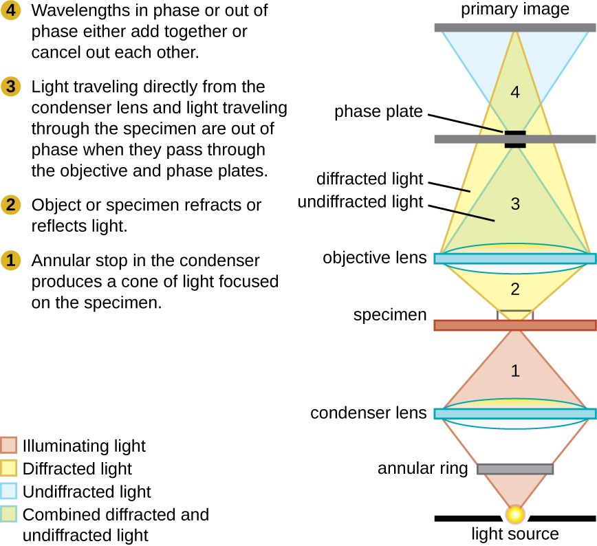
Usually the light is passed through a condenser to focus it on the specimen to get maximum illumination.
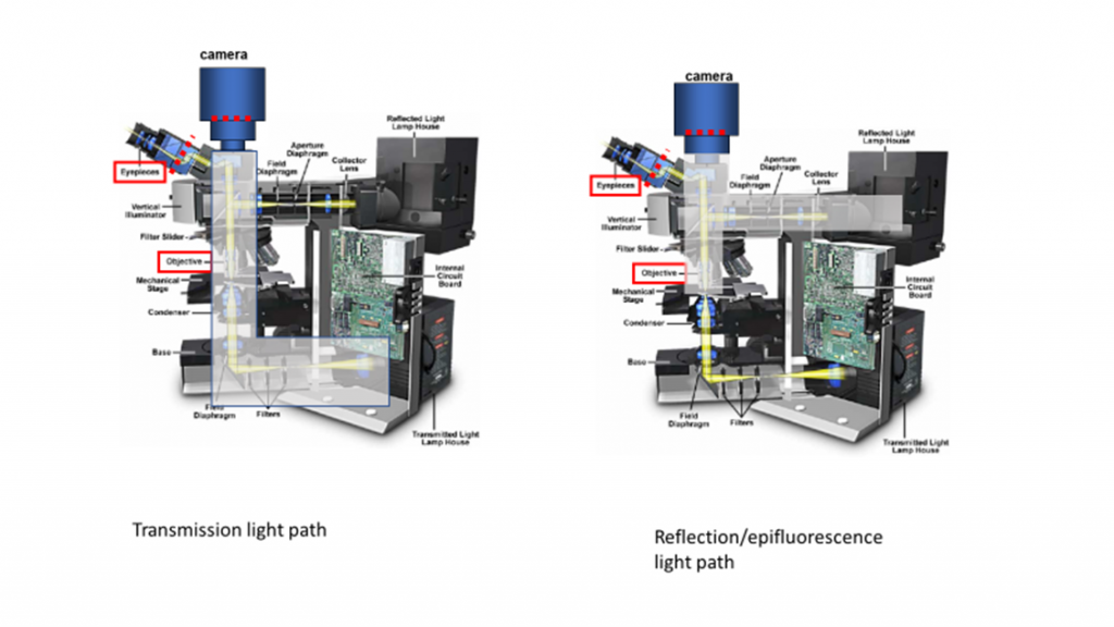
Transmitted light microscopy optical pathways.
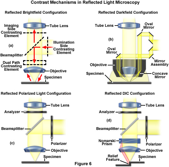
The optical pathway for reflected light begins with illuminating rays originating in the lamp housing for reflected light the upper housing in figure 2.
The optical pathway for reflected light begins with illuminating rays originating in the lamp housing for reflected light.
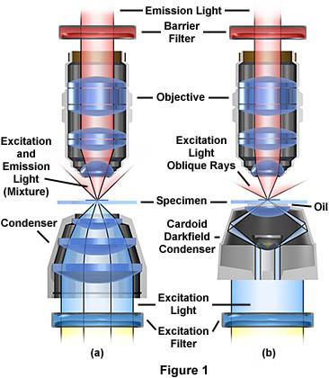
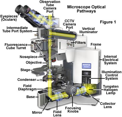
A most common light source because of its low cost and long life is the 50 or 100 watt tungsten halogen lamp as illustrated at the base of the microscope diagram in figure 1 which also details the optical pathways in a typical modern transmitted light microscope.
Background of similar intensity.
This tutorial explores the optical pathways in a transmitted light microscope.
Transmitted light microscopy is the general term used for any type of microscopy where the light is transmitted from a source on the opposite side of the specimen to the objective lens.
Instructions and a discussion about how to operate this tutorial appear immediately below the window.
The term transmitted light when used in optical microscopy refers to any imaging modality where light is passed from the illumination source on the opposite side of the specimen to the objective thus illumination is transmitted through the specimen.
Transmitted light microscopy optical pathways.
To operate the tutorial use the slider bars beneath the microscope to control the aperture of the field diaphragm.
Basic optical microscopes can be very simple although many complex.
Instructions and a discussion about how to operate this tutorial appear immediately below the window.
Optical microscopes are the oldest design of microscope and were possibly invented in their present compound form in the 17th century.
There are numerous light sources available to illuminate microscopes both for routine observation and critical photomicrography.
Today many microscope manufacturers offer advanced models that permit the user to alternate or simultaneously conduct investigations using both vertical and transmitted illumination.
Illumination of the specimen is the most important controllable variable in achieving high quality images in microscopy critical photomicrography and digital imaging.
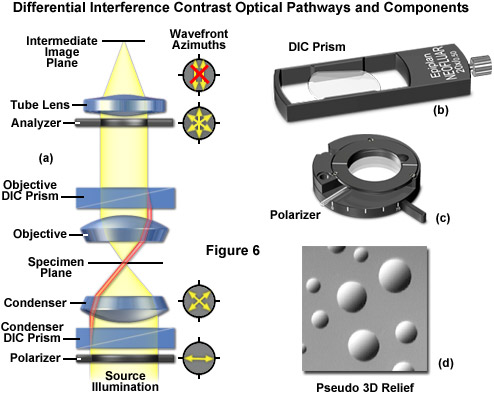
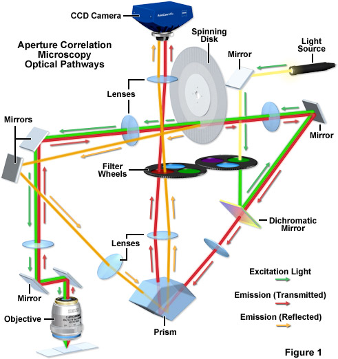
We present a new set of symmetrical ray diagrams of the optical pathways in transmitted reflected and epifluorescence light microscopes equipped with infinity corrected optics.
This tutorial explores the optical pathways in a transmitted light microscope.
The optical microscope also referred to as a light microscope is a type of microscope that commonly uses visible light and a system of lenses to generate magnified images of small objects.
To operate the tutorial use the slider bars beneath the microscope to control the aperture of the field diaphragm left slider the aperture condenser diaphragm.